Separating the light source from the instrument
In order to improve the efficiency of Desormeaux’s endoscope, efforts were undertaken to separate the light source from the reflector to avoid the many inconveniences associated with this arrangement. These shortcomings were not only inconvenient for the examining doctor but also detrimental to the well-being of the patients.
In 1873, the Viennese dermatologist Josef Grünfeld separated the light from the shaft of the endoscope and used a plane mirror for the reflection of direct sunlight or artifical light. When using artificial light he preferred petroleum light in the form of a Mitrailleuse burner, which had numerous small wicks arranged in a circle. Joseph Grünfeld designed several endoscopes for urethroscopy and cystoscopy and he is known as one of the founders of modern urethroscopy.
Source: Matthias Reuter, Hans J. Reuter, Rainer Engel: History of Endoscopy, pp 61.
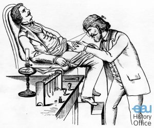 Urethroscopy according to Joseph Grünfeld. He preferred to have the patient sit on a special examination table. From the collection of the Int. Nitze-Leiter Research Society for Endoscopy.
The particular endoscope devised and used by Grünfeld successfully consisted of a large and straight metal sound that was introduced through the urethra. An external light was reflected and concentrated into the tube by a parabolic and concave mirror which the observer held in front of his eye. Grünfeld succeeded in inspecting the urethra (and with female patients also the orifices of the ureters) and in gaining also a view of the bladder wall, though limited. He was the first to catheterise the ureters under endoscopic control, to observe, fragment and remove a bladder tumor under vision, to observe endoscopically the colliculus seminalis and to describe its pathological changes. Moreover, he was able to give a detailed account of the normal and pathological aspects of the urethral mucosa by endoscopy.
Grünfeld applied endoscopy under direct vision. The reflection tubes of his endoscopes were not supported by technical aids, such as lens systems, prisms etc. for image transport. The image of the illuminated object was depicted in precisely the way one saw it through the endoscope shaft with the “naked eye”. Therefore, the image quality depended primarily on the diameter of the lumen and the length of the shaft. This is why, in this type of endoscopy, instruments with a short shaft and a lumen that was rather on the large side were preferred.
Source: Matthias Reuter, Hans J. Reuter, Rainer Engel: History of Endoscopy, pp 159-167.
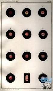 Endoscopic picture of urethral gonorrhea. In.”Formen des Harnröhrentrippers und die endoskopischen Befunde derselben”, by Josef Grünfeld, Vienna, 1877. From the collection of the Int. Nitze-Leiter Research Society for Endoscopy.